Stock solutions preparation
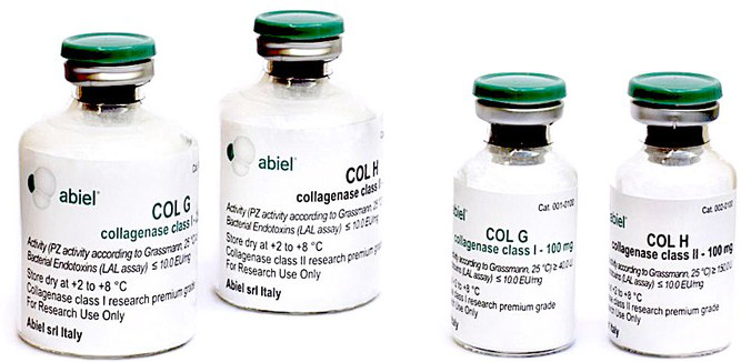
1. Dissolve COL G in 1.5 ml sterile H2O and make 3 aliquots of 500 μl (each aliquot is the Solution A) and store at -20°C
2. Dissolve COL H in 3 ml sterile H2O and make 3 aliquots of 1 ml (each aliquot is the Solution B) and store at -20°C
3. Dissolve the Thermolysin in 1.5 ml sterile H2O and make 3 aliquots of 500 μl (each aliquot is the Solution C) and store at -20°C.
Isolation of Hepatocytes from 1 rat liver
DIGESTION SOLUTION FOR 1 rat liver (weight between 180 - 220 grams):
Solubilize one aliquot of Solution A, one aliquot of Solution B in 50 ml DMEM low glucose + 1% Antibiotics (Penicillin and streptavidin) + 15 mM HEPES and put it on ice. Immediately before use add one aliquot Solution C.
WASHING SOLUTION: 100 ml of PBS solution without Ca2+ and Mg2+, containing 0.5 mM EGTA and 25 mM HEPES pH 7.4
1. Euthanize the rat (weight between 180 - 220 grams) by cervical dislocation.
2. After disinfection by alcohol and iodine solution, transfer it into a laminar flow hood and position in supine view on an inclined plane support. Using curved scissors with pliers help practicing longitudinal incision at the medial plane of the abdomen and then cut through the skin from the pelvis to the sternum.
3. Make the reversal of abdominal peritoneum muscles, exposing the viscera in order to have an easy access to the portal vein (VP) so as to be able to insert a cannula and maintain perfusion. Insert the cannula and start the perfusion with the WASHING SOLUTION with a flow rate of 2 ml/min. When the liver "bleaches" (becomes clear), cut the swollen vena cava (VC) to reduce the pressure and increase the flow rate to 9 ml/min in continuous perfusion with the WASHING SOLUTION for 10 minutes.
4. Continue perfusion with 40 ml of DIGESTION SOLUTION at the same flow rate (9 ml/min).
5. Following the perfusion, withdraw the liver from the animal and place it into a 50 ml conical tube containing 10 ml of DIGESTION SOLUTION and incubate in a thermostatic bath at 37 ° C for 10’, stirring (100 rpm).
6. Add 20 ml DMEM-low glucose + 1% Penn-Strep + 15 mM HEPES.
7. Use a 25 ml serological pipette to homogenate the digested tissue.
8. Filter through a gauze (100 mm mesh) to remove debris and undigested tissue, then centrifuge at 50g for 2’ at 4 °C.
9. Wash the pellet three times in 30 ml of DMEM-low glucose + 1% Penn-Strep + 15 mM HEPES.
10. Remove the supernatant and resuspend the hepatocytes in 10 ml DMEM/F-12 + 1% Penn-Strep.
11. Count the hepatocytes and seed into a petri dish, previously coated with rat tail type-I collagen (50 mg/ml), about 3x10⁶ cells/petri dish (7 cm diameter).
12. Incubate at 37 °C, 5% CO2, 95% O2 about 1h; then change the medium with DMEM-low glucose + 1% Penn-Strep + 10% FBS.
13. After 4-5 hours in culture (necessary time for hepatocytes adhesion) change the medium with the William’s Media E to preserve the original morphology.